Anatomy Of Chest / Man Head And Chest Anatomy Diagram With Ghost Stock Illustration 50354923 Pixta / The shape of the chest is often regarded as potential insight into a disease process, as in the case of barrel chest and respiratory dysfunction.
Anatomy Of Chest / Man Head And Chest Anatomy Diagram With Ghost Stock Illustration 50354923 Pixta / The shape of the chest is often regarded as potential insight into a disease process, as in the case of barrel chest and respiratory dysfunction.. Find out more about the individual muscles within the chest anatomy by clicking their respective links throughout this This page provides an overview of the chest muscle group. Anatomy of the thorax shari l. Plus, how to target each to make them bigger and stronger. The skeleton of the thoracic wall is formed by the twelve thoracic vertebra posteriorly,
Swensen fund for innovation in teaching. Learn about each of these muscles, their locations, functional anatomy and exercises for them. In insects, crustaceans, and the extinct trilobites, the thorax is one of the three main divisions of the creature's body, each of which is in turn composed of multiple segments. The skeleton of the thoracic wall is formed by the twelve thoracic vertebra posteriorly, An overview of the anatomy visible in a transverse computed axial tomographical image of the thorax (and part of the abdomen) performed with intravenous cont.
Figure 3 From Relevant Surgical Anatomy Of The Chest Wall Semantic Scholar from d3i71xaburhd42.cloudfront.net Anatomy of the thoracic wall. Radiology basics of chest ct anatomy with annotated coronal images and scrollable axial images to help medical students and junior doctors learning anatomy. The thoracic skeleton creates a protected space for the heart. Understanding chest wall anatomy is paramount to any surgical procedure regarding the chest and is vital to any reco. Fill out your shirt with a bigger, stronger, more powerful chest. Strictures, acute syndromes, neoplasms and vascular impressions Stability to arm and shoulder movement; The shape of the chest is often regarded as potential insight into a disease process, as in the case of barrel chest and respiratory dysfunction.
Here, we break down the anatomy of your chest muscles.
(1) the pectoralis major, and (2) the pectoralis minor. Structures to identify • heart • lungs • mediastinum • pleural space • chest wall • …everything else! It provides access to ct images in the axial plane, allowing the user to learn and review the lung anatomy interactively. The epidermis is the outermost layer that provides a protective, waterproof seal over the body. Find out more about the individual muscles within the chest anatomy by clicking their respective links throughout this The chest wall is a complex system that provides rigid protection to the vital organs such as the heart, lungs, and liver; The skeleton of the thoracic wall is formed by the twelve thoracic vertebra posteriorly, The major muscle in the chest is the pectoralis major. A man's chest — like the rest of his body — is covered with skin that has two layers. Having to do with the chest. Radiology basics of chest ct anatomy with annotated coronal images and scrollable axial images to help medical students and junior doctors learning anatomy. The chest is made up primarily of two muscles: Stability to arm and shoulder movement;
This page provides an overview of the chest muscle group. And flexibility to aid in the functional process of respiration. It spreads out like a fan and covers the rib cage like an armor plate. The chest is the area of origin for many of the body's systems as it houses organs such as the heart, esophagus, trachea, lungs, and thoracic diaphragm. Radiology basics of chest ct anatomy with annotated coronal images and scrollable axial images to help medical students and junior doctors learning anatomy.
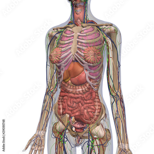
Female Anatomy Of Chest And Abdomen On White Background 2 Buy This Stock Illustration And Explore Similar Illustrations At Adobe Stock Adobe Stock from as2.ftcdn.net Here, we break down the anatomy of your chest muscles. (1) the pectoralis major, and (2) the pectoralis minor. The chest is made up primarily of two muscles: It provides a protective framework for… The pec major) is the one that commands the most real estate. The thoracic skeleton creates a protected space for the heart. Thoracic cavity, also called chest cavity, the second largest hollow space of the body.it is enclosed by the ribs, the vertebral column, and the sternum, or breastbone, and is separated from the abdominal cavity (the body's largest hollow space) by a muscular and membranous partition, the diaphragm.it contains the lungs, the middle and lower airways—the tracheobronchial tree—the heart. Definition (nci_cdisc) the anterior side of the thorax from the neck to the abdomen.
It provides a protective framework for…
The chest or thorax is the region between the neck and diaphragm that encloses organs, such as the heart, lungs, esophagus, trachea, and thoracic diaphragm. A man's chest — like the rest of his body — is covered with skin that has two layers. The pec major) is the one that commands the most real estate. Having to do with the chest. It provides a protective framework for… Chest muscles anatomy (1) pectoralis major muscle. Fill out your shirt with a bigger, stronger, more powerful chest. It provides access to ct images in the axial plane, allowing the user to learn and review the lung anatomy interactively. The chest anatomy includes the pectoralis major, pectoralis minor and the serratus anterior. Stability to arm and shoulder movement; Thoracic cavity, also called chest cavity, the second largest hollow space of the body.it is enclosed by the ribs, the vertebral column, and the sternum, or breastbone, and is separated from the abdominal cavity (the body's largest hollow space) by a muscular and membranous partition, the diaphragm.it contains the lungs, the middle and lower airways—the tracheobronchial tree—the heart. Studied the anatomy of the breast, its topography, innervation, vascularization and lymphatic drainage, and correlated the anatomical data with the classification of lymph node groups that is frequently utilized by mastologists. Computed tomography (ct) of the chest can detect pathology that may not show up on a conventional chest radiograph (1).
The chest anatomy includes the pectoralis major, pectoralis minor and the serratus anterior. In insects, crustaceans, and the extinct trilobites, the thorax is one of the three main divisions of the creature's body, each of which is in turn composed of multiple segments. About the 6th week, the somites differentiate into the sclerotomes and the dermatomyotomes. Structures to identify • heart • lungs • mediastinum • pleural space • chest wall • …everything else! Download my two educational text books for free using this link:
6 494 Anatomy Chest Photos Free Royalty Free Stock Photos From Dreamstime from thumbs.dreamstime.com This page provides an overview of the chest muscle group. The epidermis is the outermost layer that provides a protective, waterproof seal over the body. In insects, crustaceans, and the extinct trilobites, the thorax is one of the three main divisions of the creature's body, each of which is in turn composed of multiple segments. Other important structures, such as the pleura, only become visible when abnormal, and some are not visible at all, such as the phrenic nerve. The chest wall is a complex system that provides rigid protection to the vital organs such as the heart, lungs, and liver; It is important to remember the position and orientation of the heart when placing a stethoscope on the chest of a patient and listening for heart sounds, and also when looking at images taken from a midsagittal perspective. The skeleton of the thoracic wall is formed by the twelve thoracic vertebra posteriorly, This atlas is a comprehensive and affordable learning tool for medical students and residents and especially for radiologists and pneumologists.
Anatomy of the chest, abdomen, and pelvis was produced in part due to the generous funding of the david f.
Other important structures, such as the pleura, only become visible when abnormal, and some are not visible at all, such as the phrenic nerve. It provides access to ct images in the axial plane, allowing the user to learn and review the lung anatomy interactively. An overview of the anatomy visible in a transverse computed axial tomographical image of the thorax (and part of the abdomen) performed with intravenous cont. The circulatory system does most of its. The shape of the chest is often regarded as potential insight into a disease process, as in the case of barrel chest and respiratory dysfunction. Thoracic cavity, also called chest cavity, the second largest hollow space of the body.it is enclosed by the ribs, the vertebral column, and the sternum, or breastbone, and is separated from the abdominal cavity (the body's largest hollow space) by a muscular and membranous partition, the diaphragm.it contains the lungs, the middle and lower airways—the tracheobronchial tree—the heart. Anatomy of the thorax, heart, abdomen and pelvis recommended text gray's anatomy for students. Applied anatomy of the chest wall and mediastinum petros mirilas michael e. It provides a protective framework for… Definition (nci_cdisc) the anterior side of the thorax from the neck to the abdomen. Stability to arm and shoulder movement; (1) the pectoralis major, and (2) the pectoralis minor. Computed tomography (ct) of the chest can detect pathology that may not show up on a conventional chest radiograph (1).